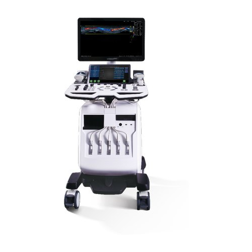
VINNO’s RF ultrasound platform, with adjustable imaging parameters, allow users to obtain outstanding images
Features
- The RF platform ensures zero loss of imaging data and improved fine detail, with enhanced image contrast and edge sharpness
- State-of-the art multi-channel platform provides superior image resolution and penetration
- Advanced RF-based post-processing algorithms guarantee users a high quality image
- Offers a wide variety of features and tools to aid diagnostics
- User-friendly interface and fast workflow offers improved efficience.
Technical Features
Innovative RF platform: a world first.
VINNO’s unique RF platform, the first of its kind, removes the need for the hardware pre-processing and demodulation of traditional ultrasound platforms. The whole signal is used for image-processing, which allows up to 40 times more data to be retained in comparison with conventional ultrasound techniques. This means that more
accurate data is available to the clinician for post-processing and ensures superior image quality in terms of resolution and contrast. The platform also has a wide frequency range which can support probes from 1-25MHz.
VTissue Tissue signature image
VTissue automatically compensates for variations in the speed of sound between different tissues to enhance imaging throughout the body.
Excellent 3D/4D Capabilities
The RF platform provides accurate volumetric image-processing alongside world-class convex and endocavity probes. This allows a high quality image for obstetric and gynaecological applications.
Pure Wave probe technology
Pure wave (single crystal) probe technology increases bandwidth and signal sensitivity in order to provide improved penetration and colour sensitivity for cardiac and abdominal applications.
Vspeckle – speckle reduction image
Speckle reduction technology utilizes automatic structure detection to eliminate noise artefacts and to provide a more accurate image of tissue.
High quality 3D/4D volumetric imaging
High quality 3D/4D volumetric imaging can be used to show the flow of a contrast bubble agent through the oviduct to check fallopian tube patency.
Smart 3D/4D touch-screen operation mode


